1. Introduction
Intestinal obstruction is a leading cause of neonatal surgical admission, with an incidence of approximately 1 per 2,000 live births. [1] Favourable outcome depend heavily on early diagnosis and transfer to specialized tertiary centres. [2] Early surgical intervention is vital to maintain bowel perfusion and avoid devastating consequences such as gangrene, perforation (pneumoperitoneum), or long-term short bowel syndrome. [3] While these infants require the expertise of trained pediatric surgeons and advanced Neonatal Intensive Care Units (NICUs), such resources remain scarce in many regions. Prognosis is further complicated by factors like premature birth, delayed clinical presentation, and co-existing congenital anomalies. [3]
The etiology of neonatal obstruction is diverse, though many cases are now suspected before birth. Prenatal ultrasound indicators such as polyhydramnios, a "double-bubble" sign suggesting duodenal atresia, or non-visualisation of stomach bubble in esophageal atresia often provide the first clues. Other intrauterine warning signs include presence of echogenic bowel, calcifications (intra-abdominal or intra-luminal), or bowel wall thickening exceeding 3mm and associated congenital anomalies.[4]. Postnatally, an abdominal X-ray serves as the primary diagnostic tool. The number of dilated bowel loops often dictates the next step. In proximal intestinal obstruction, fewer than four dilated loops are seen, and is typically evaluated further via an upper gastrointestinal (UGI) contrast series. While, distal intestinal obstruction is suggested by four or more dilated loops and it generally requires a contrast enema to differentiate other potential causes.
Newborns typically present with a triad of symptoms: bilious vomiting, abdominal swelling, and a failure to pass meconium within the first 24–48 hours. Initial management focuses on stabilization followed by definitive management. The management protocol includes:
a) Early Bowel Decompression: Insertion of a large-bore nasogastric tube.
b) Supportive Care: Stopping oral intake, initiating intravenous fluids and antibiotics, and strictly maintaining body temperature (especially in preterm infants).
c) Intensive Monitoring: Continuous assessment of hemodynamic status, glucose, electrolytes, and acid-base balance in a NICU setting ensures the infant is stable for further surgical correction. [5]
d) Definitive Management: As per diagnosis, the neonate is investigated and managed accordingly. The specific procedure depends on the etiology (e.g., resection and anastomosis for atresia, Ladd's procedure for malrotation).
In this chapter, an overview of the common causes of neonatal intestinal obstruction has been studied. Moreover, it also highlights an approach to evaluate and further manage the neonates with intestinal obstruction in emergency.
2. Embryological Pathophysiology: The primitive gut tube differentiates from the yolk sac around the fourth gestational week, organizing into three distinct anatomical zones—foregut, midgut, and hindgut which is defined by their specific arterial supply. [6]
Foregut Anomalies (Celiac Axis)
Malformations in this region typically manifest as proximal obstructions.
Duodenal Atresia: This condition stems from a failure in the "recanalization" phase. Between the 8th and 10th gestational weeks, the duodenal lumen which is temporarily a solid cord of epithelium fails to vacuolize and restore patency.
Annular Pancreas: This results from a rotational error of the ventral pancreatic bud. Instead of migrating dorsally to fuse with the dorsal bud, it remains fixed, forming a ring of pancreatic tissue that encircles and constricts the second portion of the duodenum.
Midgut Anomalies (Superior Mesenteric Artery)
Disorders here often involve rotational mechanics or vascular integrity.
Malrotation & Volvulus: Normal intestinal rotation involves a 270-degree counter-clockwise turn around the superior mesenteric artery. Arrested rotation leads to a narrow mesenteric base (Ladd's bands), predisposing the bowel to twist (volvulus) and compromise blood flow. Jejunoileal Atresia: Unlike duodenal atresia, these lesions are typically acquired in utero due to a vascular accident (e.g., fetal intussusception or volvulus) that causes ischemic necrosis and subsequent resorption of the affected bowel segment.
Hindgut Anomalies (Inferior Mesenteric Artery) Pathology in the distal gut is frequently linked to neuro-migration or septation errors.
Hirschsprung’s Disease: This neurocristopathy is caused by the incomplete craniocaudal migration of neural crest cells. Failure of these cells to reach the distal rectum (typically by week 12) results in an aganglionic segment that cannot relax, causing functional obstruction. The RET proto-oncogene is a key genetic driver.
Anorectal Malformations: These defects arise between the 4th and 8th weeks when the urorectal septum fails to completely partition the cloaca into the urogenital sinus and anorectum. This often leaves a fistula connecting the rectum to the urinary or genital tract. [7] Systemic Etiology Meconium Ileus: Predominantly associated with Cystic Fibrosis (CF), this condition is driven by CFTR gene dysfunction. Meconium ileus is an intestinal obstruction at the level of the terminal ileum that occurs when inspissated meconium obstructs the intestinal lumen.
Meconium ileus is the earliest sign of cystic fibrosis (CF), an autosomal recessive condition that is characterized by abnormalities in cellular membrane physiology and chloride ion transport which causes progressive respiratory failure, derangements in cellular secretory patterns, and diminished mucosal motility. Around 15% of newborns with CF present with meconium ileus.[ 8]
3. Etiopathogenesis
Neonatal intestinal obstruction is clinically categorized as "high" or "low" based on the count of dilated bowel loops visible on initial X-rays. [9] A "high" obstruction—located proximal to the ileum—typically presents with three or fewer dilated loops (often involving the stomach, duodenum, or jejunum). Conversely, a "low" obstruction involves the distal ileum or colon and is characterized by more than three dilated small-bowel loops. [Table-1] While certain high obstructions (like duodenal atresia) may lead directly to surgery, an upper GI series is the standard follow-up for further evaluation. For suspected low obstructions, a contrast enema is typically the next step to identify the specific site of the obstruction.
Table 1:
Causes of Neonatal Intestinal Obstruction
High intestinal obstruction Esophageal Atresia Gastric Atresia, Pyloric Atresia Duodenal atresia Duodenal stenosis (with annular pancreas)
Duodenal web Malrotation Jejunal atresia and stenosis Low intestinal obstruction Ileal atresia Meconium ileus Functional immaturity of the colon Hirschsprung disease Colonic atresia Anal atresia and anorectal malformations Miscellaneous Postoperative adhesions Para-duodenal hernias Congenital Bands Pseudo-obstruction (NEC, Hypothyroidism etc.)
4. Clinical Presentation and Initial Assessment
Neonatal intestinal obstruction typically presents with a triad of symptoms: bilious emesis, abdominal swelling, and failure or delay in passing meconium. [10] When a neonate presents with these signs in a primary care or peripheral setting, the clinician’s primary objectives are to:
Distinguish between functional (physiological) and mechanical (pathological) obstruction. Determine if the distension requires medical management or surgical intervention.
Identify immediate red flags and stabilize the infant.
Initiate essential diagnostic workup and early referral for surgical consultation.
Following a standardized diagnostic algorithm [Figure 1] can help streamline the identification and management of these cases.
Physical Examination Priorities
A systematic physical exam for a suspected obstruction should evaluate: Vital Signs: Assess overall activity, core temperature, and signs of peripheral perfusion (shock/dehydration).
Gastric Output: Note the colour and consistency of nasogastric aspirate or vomit (specifically looking for bile).
Systemic Status: Check for complications like sepsis or signs of Necrotizing Enterocolitis (NEC).
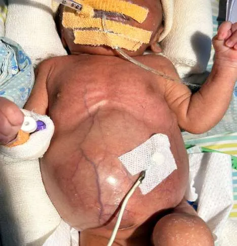
Abdominal Inspection: Look for visible venous patterns, redness (erythema), or a tense, glossy abdominal wall.
Anatomic Check: Inspect the genitalia and hernial orifices for incarceration.
Rectal Patency: Perform a cautious digital or probe examination to check for anal patency and the character of meconium triggered by stimulation.
Comorbidities: Screen for any obvious congenital anomalies.
In proximal obstructions—such as duodenal stenosis or atresia, or midgut volvulus—the abdomen often appears flat or only minimally distention is noted. [11] Despite the lack of physical signs, conditions like midgut volvulus are surgical emergencies because they can lead to rapid and extensive bowel gangrene.
Key diagnostic principles include
Imaging Protocol: If obstruction is suspected, an upright abdominal X-ray (covering the area from the neck to the knees) is the first step. The resulting gas patterns are often definitive for diagnosis.
The "Golden Rule": Any previously healthy infant presenting with green (bilious) vomit must be treated as having a midgut volvulus until a surgeon proves otherwise.
Prenatal Indicators: A history of maternal polyhydramnios on ultrasound is a strong indicator of a potential congenital intestinal obstruction or atresia.
Contrast Studies: An upper GI series is typically used for suspected partial proximal obstruction (like malrotation), while a contrast enema is the preferred tool for evaluating distal bowel obstructions such as Hirschsprung’s disease, ileal atresia, or meconium ileus.[12, 13] The underlying causes of these obstructions vary significantly, and accurate radiological assessment is essential for distinguishing between cases requiring medical versus surgical management. The etiopathogenesis of neonatal intestinal obstruction is varied depending on the primary cause of obstruction. (Table 2).
Table 2: Differential diagnosis of Neonatal Intestinal Obstruction
Medical causes Neonatal Sepsis Metabolic ileus, Maternal drug induced Maternal hypothyroidism, Maternal diabetes mellitus Neonatal small left colon syndrome, Meconium plug syndrome Neonatal Necrotizing Enterocolitis (Uncomplicated)
Surgical causes Duodenal atresia or stenosis, Malrotation of gut with midgut volvulus, Intestinal atresia (Jejunoileal/ Colonic)
Meconium Ileus, Meconium Peritonitis Congenital band obstruction (Meckel’s band etc.)
Neonatal Necrotizing enterocolitis (Complicated)
Hirschsprung’s disease Anorectal Malformation
Initial Neonatal Assessment
The infant's overall activity and general state often provide the first clues toward a diagnosis. Clinicians should begin by taking a detailed history to exclude common non-pathological issues, such as poor feeding techniques, inadequate burping, or drug-induced ileus from maternal medications. However, high-alert symptoms like bilious vomiting or haematochezia (bloody stools) must be treated with urgency. Systemic instabilities—including electrolyte imbalances, circulatory collapse, or clotting disorders—are more frequently associated with medical conditions such as Necrotizing Enterocolitis (NEC). [6, 14]
Radiological Interpretation
Plain abdominal X-rays are the cornerstone of diagnosis, focusing on the distribution and paucity of intestinal gas:
- Gas Distribution: The clinician looks for a lack of distal gas or an uneven gas pattern, which points toward an intestinal obstruction below.
- Positioning: A supine view is best for estimating the anatomical level of the obstruction based on how far the gas has travelled. An upright (erect) view confirms the diagnosis, especially if multiple air-fluid levels are visible.
- Complications: The presence of a pneumoperitoneum (free air under diaphragm) indicates a perforation, often resulting from atresia, meconium ileus, or Hirschsprung’s disease. [14, 15]
- Medical vs. Surgical: In non-surgical cases like neonatal sepsis, the X-ray typically shows uniformly dilated loops without the significant air-fluid levels or free air seen in mechanical obstructions.
Differential Diagnosis by Imaging Findings
Proximal Obstructions
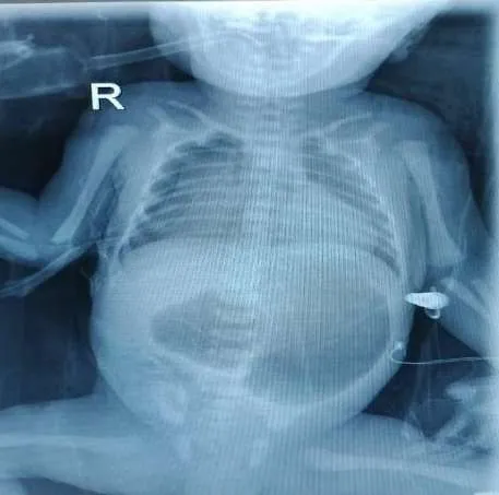
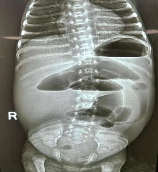
Duodenal Atresia: Identified by the "double bubble" appearance and a total lack of gas in the distal bowel. [Figure 2(a)]
Duodenal Stenosis: Similar "double bubble" sign, but with small amounts of gas visible distally.
Malrotation with Midgut Volvulus: Characterized by gastric and duodenal dilation with minimal distal gas; in advanced cases, the abdomen may appear entirely gasless.
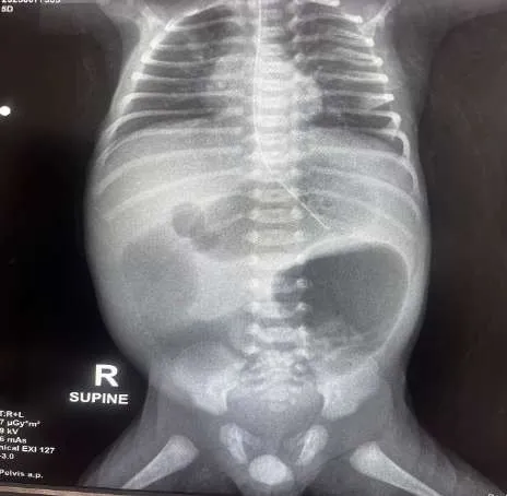
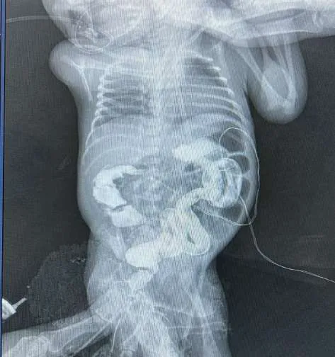
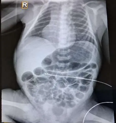
Jejunoileal Atresia: Typically presents as a "triple bubble" sign or several air-fluid levels, depending on the specific site of the atresia with no distal gas. [Figure 2(b)]
Distal Obstructions
Meconium Ileus: Shows dilated small bowel loops without air-fluid levels, often accompanied by a granular "soap bubble" (Neuhauser) appearance.
Meconium Peritonitis: Note for intra-abdominal calcifications on right sided lower abdomen on supine radiographs.
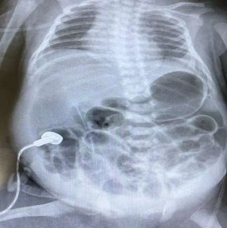
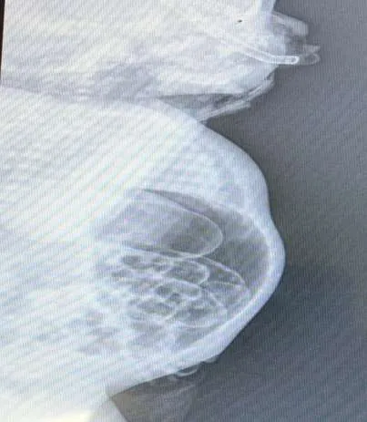
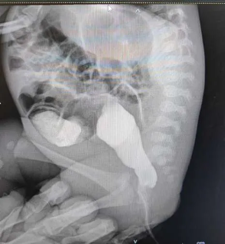
Distal Small Bowel involvement: For low obstruction, an intestinal perforation will show a "football sign" on supine radiograph indicating a large pneumoperitoneum. [Figure 3(a)] while lateral or erect abdominal X-rays will show free gas in abdomen. [Figure 3(b)] [15, 16] A contrast enema is necessary to differentiate between ileal atresia (shows a "microcolon"), meconium ileus, or total colonic Hirschsprung’s Disease (HD) patients without perforation. [16, 17]
Colonic & Functional Issues
Hirschsprung’s Disease (HD): Plain films show multiple air fluid levels with dilated loops and absent distal rectal gas. [Figure 4] Contrast studies reveal a narrow rectum with a cone-shaped transition zone. [Figure 5(a)] The "question mark" colon is a hallmark of the total colonic variant. [14-16]
Small Left Colon Syndrome: Presents similarly to HD on X-ray, but contrast studies show a narrow left colon that widens at the splenic flexure without a true transition zone. [Figure 5(b)] Necrotizing Enterocolitis (NEC): Usually seen in stressed pre-term infants; features on plain radiograph include pneumatosis intestinalis (gas in the bowel wall). [17] Advanced signs include portal venous gas, a "fixed loop," or pneumoperitoneum. [Figure 6(a&b)]
Sepsis-Related Ileus: Distinguished from surgical cases by uniform bowel dilation, an absence of discrete air-fluid levels, and the presence of distal gas.
5. Management
General Preoperative Care
Across all forms of neonatal obstruction, initial stabilization is mandatory. This involves aggressive fluid resuscitation, continuous nasogastric decompression, and the administration of prophylactic broad-spectrum antibiotics. [18]
Pathology-Specific Protocols
Duodenal Atresia: As this is often part of a spectrum of midline defects, infants must be screened for Trisomy 21. A comprehensive anomaly survey is required, including echocardiography, renal and cranial ultrasounds, and vertebral X-rays.
Jejunoileal Atresia: During surgery, the patency of the distal bowel must be verified. Surgeons typically flush the distal lumen with warm saline to ensure there are no secondary "downstream" atresia before reaching the rectum.
Malrotation and Volvulus: Ultrasound is now a preferred primary diagnostic tool.
Malrotation is suspected if the SMA/SMV relationship is inverted (SMA to the right of the SMV). A "whirlpool sign" on Doppler is a surgical emergency, indicating active volvulus. Meconium Ileus: Water-soluble contrast enemas are now favoured over traditional gastrograffin contrast to clear meconium impactions, provided the contrast reaches the terminal ileum. To avoid complications like hypovolemic shock or perforation (risk 3–10%), hyperosmolar solutions should be used with extreme caution. N-acetylcysteine via nasogastric tube can further assist in softening the meconium. Meconium Plug Syndrome: Many cases resolve quickly with gentle rectal irrigation using lukewarm normal saline.
Hirschsprung’s Disease (HD): Initial relief is achieved through rectal washouts. Diagnosis is confirmed with rectal suction biopsy or full-thickness biopsy if specialized histochemistry is unavailable. A contrast enema is used to map the transition zone and disease extent.
Anorectal Malformations: Evaluation is delayed for at least eighteen hours to allow swallowed air to reach the distal rectum. A cross-table prone X-ray is then used to differentiate between high and low lesions.
Surgical Management Strategies
Duodenal Atresia: Obstruction is typically bypassed using a duodeno-duodenostomy, often performed in a "diamond-shaped" or side-to-side configuration.
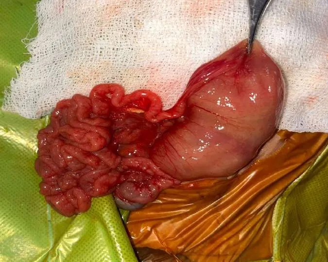
Malrotation and Volvulus: This is a critical surgical emergency; any delay increases the risk of total bowel loss. If the intestine is viable, it is detorted counter-clockwise. This is followed by a Ladd’s procedure, which involves mobilizing the mesentery to prevent recurrence. Jejunoileal Atresia: Management focuses on resecting the most dilated proximal segment followed by a primary end to end anastomosis. [Figure 7] While a diverting stoma is avoided, when possible, the proximal bowel may be tapered to improve motility. Preserving the ileocecal valve is a priority to prevent bacterial overgrowth and malabsorption.
Meconium Ileus & Peritonitis
Complicated: If X-rays show calcification or the infant is born with a red, swollen abdomen (indicating in-utero perforation), a laparotomy is required. Surgeons drain any pseudocysts and typically convert the perforation site into an enterostomy.
Uncomplicated: The bowel may be cleared via irrigation through an enterostomy or T-tube. T-tubes allow for postoperative contrast checks and can be removed once patency is confirmed, allowing the site to close without further surgery.
Hirschsprung’s Disease (HD): Unless complicated by enterocolitis, the goal is surgical correction. While a levelling colostomy at the transition zone was traditional, many cases are now managed with primary pull-through procedures (open or laparoscopic). Recent trends favor minimally invasive techniques, such as the trans-anal endorectal pull-through.
Anorectal Malformations
Low Lesions: Lesions with a perianal fistula are generally corrected with a primary anoplasty. High Lesions: If the fistula connects to the urinary tract or vagina, an initial diverting colostomy is usually performed. Definitive reconstruction typically involves a posterior sagittal anorectoplasty (PSARP) or a laparoscopic-assisted pull-through to precisely position the rectum within the sphincter complex. [19]
6. Postoperative Support and Interdisciplinary Care
The improved survival rates for neonates with intestinal obstructions are largely due to advances in Neonatal Intensive Care Unit (NICU) support. Critical components of recovery include meticulous fluid management, total parenteral nutrition (TPN), and specialized respiratory care. In regions where TPN is inaccessible, trans-anastomotic feeding tubes are often utilized to initiate early enteral nutrition, though outcomes vary.
Condition-Specific Recovery
- Anorectal Malformations: Following a posterior sagittal anorectoplasty, a regimen of progressive anal dilation typically begins two weeks post-surgery to maintain patency and prevent strictures.
- Systemic Follow-up: Long-term management often extends beyond the initial surgical recovery. For conditions linked to systemic disorders —such as cystic fibrosis (associated with meconium ileus) or Trisomy 21 (associated with duodenal atresia)—early and ongoing consultation with specialists is essential for the child's overall development.
Collaborative Workflow
The cornerstone of successful treatment is the seamless coordination between neonatologists and paediatric surgeons. This partnership ensures that diagnostic steps are streamlined, and life-saving interventions are delivered without delay.
7. Potential Postoperative Complications
- Treatment-Related Risks: Infants requiring prolonged intensive care are at risk for complications stemming from life-support measures. Total Parenteral Nutrition (TPN) can lead to cholestasis or hyperalimentation -induced hepatitis. Additionally, central venous access carries risks such as catheter-related sepsis, pneumothorax, or rare catheter emboli.
- Mechanical and Structural Issues:
Strictures and Adhesions: Post-surgical narrowing (anastomotic stricture) or the development of adhesions can occur after any abdominal surgery, often exacerbated by a history of peritonitis. Research indicates that nearly 10% of pediatric intestinal surgery patients develop adhesions at the operative site, with an additional 5% occurring elsewhere in the abdomen. [20]
Dysmotility: Even after a surgical obstruction is cleared, the formerly dilated proximal bowel may exhibit poor peristalsis, leading to chronic motility issues.
Malabsorption and Short Bowel Syndrome (SBS): SBS occurs when the remaining intestinal length is insufficient for nutrient absorption. A full-term infant typically has about 250 cm of small bowel; a minimum of roughly 75 cm is generally needed for adequate function. Resecting more than 60% of the intestine or losing the ileocecal valve significantly increases the risk of malabsorption and bacterial overgrowth. Advanced Management of SBS include: Medical and Nutritional: Probiotics may help stabilize intestinal flora and reduce failure to thrive. Hormonal therapies are sometimes used to encourage bowel adaptation.
Surgical Interventions: Procedures like the Serial Transverse Enteroplasty (STEP) can increase functional bowel length.
Transplantation: In severe cases, small bowel or multi-visceral transplantation is an option, though it remains a highly specialized procedure available only at select global centres.
8. Prognosis and Clinical Outcomes
The survival and long-term success for infants with intestinal obstruction are determined by several critical factors: the speed of diagnosis, the quality of preoperative stabilization, and the technical skill of the surgical and anaesthetic teams. Above all, the availability of specialized Neonatal Intensive Care (NICU) is the primary determinant of a positive outcome; without this specialised postoperative support, the prognosis remains guarded.
While neonatal intestinal obstruction is a life-threatening emergency, the overall outlook is generally optimistic when surgical correction is performed promptly. However, final outcomes are heavily influenced by the specific cause of the obstruction, the timing of the intervention, and the level of medical resources available at the treating facility.
Overall Prognosis and Survival Rates
- Developed Countries: Survival is high, with mortality often below 5% due to specialized neonatal intensive care units (NICUs) and early multidisciplinary management.
- Developing Countries: Mortality rates are higher, ranging from 9% to over 40% in various studies. Factors contributing to this include late presentation, inadequate equipment, and limited access to pediatric surgical services.
Key Outcomes and Complications
- Common Post-Surgical Complications:
- Sepsis/Septicaemia: The most frequent complication and leading cause of death.
- Wound Infection: Observed in 5% to 16% of cases.
- Anastomotic Leak: Occurs in roughly 2% to 5% of neonates, sometimes requiring re-exploration.
- Pneumonitis/Respiratory Failure: Often associated with aspiration or post-operative recovery.
- Long-Term Outlook:
- Short Gut Syndrome: A risk if a significant portion of the bowel is necrotic or resected (e.g., in midgut volvulus).
- Normal Function: Most survivors of atresia achieve relatively normal bowel function.
- Persistent Issues: Some conditions, like Hirschsprung’s disease, may lead to chronic constipation or enterocolitis even after repair.
Predictors of Outcome
The following factors significantly influence mortality and morbidity:
Timing of Intervention: Delays in diagnosis and surgery are major risk factors for bowel ischemia and death, especially in midgut volvulus.
Birth Weight and Gestation: Low birth weight (<2.5 kg) and prematurity are strongly associated with poorer survival and higher complication rates.
Associated Anomalies: The presence of other congenital defects (e.g., cardiac defects, Down syndrome) increases mortality risk. Pre-operative State: Factors like sepsis at admission, metabolic acidosis, and the need for mechanical ventilation are predictive of adverse outcomes.
In summary, intestinal obstructions represent the most frequent surgical emergencies in the neonatal period, making prompt and precise identification vital. Familiarity with the distinct radiographic signs of these conditions allows clinicians to either confirm a diagnosis immediately or determine the necessary next steps for further imaging. When initial X-rays indicate a proximal intestinal obstruction, an upper gastrointestinal series is the standard secondary study. However, for certain classic presentations such as the "double bubble" seen in duodenal atresia and the diagnosis is often sufficiently clear to proceed directly to surgery. In cases where imaging suggests a distal obstruction, a contrast enema remains the definitive diagnostic tool for further investigation. Management of neonatal intestinal obstruction needs specialized care in a tertiary care with surgical NICU facility. Initial stabilization followed by definitive treatment prevents postoperative complications and may lead to early recovery with better outcomes in these neonates.
Key Takeaways for Clinical Practice
- Urgent Surgical Status: Obstructions in the intestinal tract represent one of the most frequent surgical emergencies in the newborn period.
- Importance of Specialized Care: Patient survival is heavily dependent on rapid identification and prompt transfer to a facility equipped with specialized pediatric surgical and neonatal intensive care (NICU) services.
- Classic Diagnostic Triad: Clinicians should maintain a high index of suspicion when an infant presents with bilious vomiting, varying levels of abdominal distension, and a failure to pass meconium.
- Diagnostic Pathway: Initial evaluation begins with plain abdominal X-rays, followed by contrast imaging or histopathological tests as needed to pinpoint the underlying cause.
- Risk Mitigation: Severe complications such as midgut volvulus, bowel gangrene, perforation (pneumoperitoneum), or aspiration pneumonia require immediate stabilization through aggressive resuscitation and rapid transport.
- Prognostic Factors: Overall outcomes are directly linked to the availability of modern medical resources and the specific expertise of the neonatal care team.
References
- Juang D, Snyder CL. Neonatal bowel obstruction. Surg Clin North Am. 2012;92:685-711. doi:10.1016/j.suc.2012.03.008.
- Farrokhkhani P, Farhadi R, Ala S, Mousavi SA. Etiology and Outcome of Intestinal Obstruction in Neonates: A 5-Year Investigation of Admitted Cases From a Tertiary Neonatal Intensive Care Unit in Northern Iran. Clin Med Insights Pediatr. 2023;17:11795565231196771. doi:10.1177/11795565231196771.
- Otim P, Elobu EA, Mbiine R, Kakembo N, Komakech D. The etiological spectrum of bowel obstruction and early postoperative outcome among neonates at a tertiary hospital in Uganda. World J Pediatr Surg. 2022;5:e000377. doi:10.1136/wjps-2021-000377.
- Desoky SM, Kylat RI, Udayasankar U, Gilbertson-Dahdal D. Managing neonatal bowel obstruction: clinical perspectives. Res Rep Neonatol. 2018;8:19-32. doi:10.2147/RRN.S125760.
- Verma A, Rattan KN, Yadav R. Neonatal intestinal obstruction: a 15 year experience in a tertiary care hospital. J Clin Diagn Res. 2016;10:10 -13. doi:10.7860/JCDR/2016/17204.7268.
- Vinocur DN, Lee EY, Eisenberg RL. Neonatal intestinal obstruction. AJR Am J Roentgenol. 2012;198(1):W1–W10. doi:10.2214/AJR.11.6931.
- 7. Ludwig K, De Bartolo D, Salerno A, Ingravallo G, Cazzato G, Giacometti C, Dall'Igna P. Congenital anomalies of the tubular gastrointestinal tract. Pathologica. 2022 Feb;114(1):40-54. doi:10.32074/1591-951X-553.
- 8. Vargas MG, Miguel-Sardaneta ML, Rosas-Tellez M, Pereira-Reyes D, Justo-Janeiro JM. Neonatal intestinal obstruction syndrome. Pediatr Ann. 2018 May 1. 47 (5):e220-5. doi:10.3928/19382359-20180425-02.
- FS Alatas. Neonatal Gastrointestinal Emergencies. Arch Pediatr Gastr Hepatol Nutr. 2024;3(1):41-57. doi:10.58427/apghn.3.1.2024.41-57.
- Sarma VP et al. The approach to a neonate with suspected intestinal obstruction: the pediatric surgical perspective. Int Surg J. 2019 Nov;6(11):4198-4202. doi:10.18203/2349- 2902.isj20195156.
- Carroll AG, Kavanagh RG, Ni Leidhin C, Cullinan NM, Lavelle LP, Malone DE.
- Comparative effectiveness of imaging modalities for the diagnosis of intestinal obstruction in neonates and infants: a critically appraised topic. Acad Radiol. 2016;23(5):559-68. doi:10.1016/j.acra.2015.12.015.
- Reid JR. Practical imaging approach to bowel obstruction in neonates: a review and update. Semin Roentgenol. 2012;47(1):21-31. doi:10.1053/j.ro.2011.07.003.
- Schulman M H. Imaging of neonatal gastrointestinal obstruction. Radiol Clin North Am. 1999;37:1163-86. doi:10.1016/s0033-8389(05)70255-4.
- Buonomo C. Neonatal gastrointestinal emergencies. Radiol Clin North Am. 1997;35:845-
- doi:10.1016/S0033-8389(22)00570-3.
- Berrocal T, Lamas M, Gutieerrez J, Torres I, Prieto C, del Hoyo ML. Congenital anomalies of the small intestine, colon, and rectum. Radiographics. 1999;19:1219 -36. doi:10.1148/radiographics.19.5.g99se041219.
- Kiely EM. Meconium ileus. In: Puri P, editor. Newborn surgery, 4th ed. Boca Raton: CRC Press; 2017. p. 705-712. doi:10.1201/b13479-112.
- Dillon PW, Cilley RE. Newborn surgical emergencies. Gastrointestinal anomalies, abdominal wall defects. Pediatr Clini North Am. 1993;40(6):1289-314. doi:10.1016/s0031- 3955(16)38662-x.
- Sidler D, Debrew M, Lakhoo K. Neonatal Intestinal Obstruction. In: Ameh, E.A., Bickler, S.W., Lakhoo, K., Nwomeh, B.C., Poenaru, D, editors. Pediatric Surgery. Springer, Cham. 2020, 645-654. doi:10.1007/978-3-030-41724-6_61.
- Peña A, Levitt M. Anorectal malformations: experience with the posterior sagittal approach. In: Stringer MD, Oldham KT, Mouriquand PDE, editors. Pediatric surgery and urology: long-term outcomes. 2nd ed. Cambridge: Cambridge University Press; 2006. p. 376-
- doi:10.1017/CBO9780511545757.032.
- Stöss C, Nitsche U, Bader FG, et al. Small bowel obstruction after pediatric abdominal surgery: a systematic review. J Pediatr Surg. 2017 Nov;52(11):1710 -1715. doi:10.1016/j.jpedsurg.2017.03.064
Figure 1: Algorithm for diagnosis of Neonatal Intestinal Obstruction
Place
   Suspicion of obstruction                     Will            No             Consider:
                             nasogastric
                                             Nasogastric
• Vomiting                   tube and
                                              tube pass?
                             resuscitate                                    •Esophageal
• Abdominal distention
                                                                               atresia or web
• Failure to pass meconium
• Abnormal anus- Yes/No
                                          Yes
                                                                No             Consider:
                                             Is Anus patent
                                              & normally
                                                                            •Imperforate
                                              positioned?
                                                                               anus
                                        Yes
                                                                                Consider:
                        Obtain plain radiograph
   Bilious Emesis
                                                                 No          • Duodenal
                                             Is air seen in
                                                                                atresia or web
                                             small intestine
                                                                                or Annular
                                             on plain
                                                                                Pancreas
                                             X‑ray?
                                                                             • Malrotation
                                                                                with volvulus
 The presence of                         Yes
                                                                             •Pyloric stenosis
 bilious emesis       Bowel gas pattern
 demands              more consistent with                        Bowel gas pattern
 investigation to     Proximal Obstruction                        more consistent with
 rule out                                                         Distal Obstruction
                      Laparotomy or Upper
 Malrotation with                                              Contrast Enema (Upper
                      GI series
 Volvulus                                                      GI may be necessary to
                                                               rule out malrotation)

                           Consider:
                                                                      Consider:
                         • Malrotation
                            with volvulus *
                                                                Hirschsprung disease#
                         • Jejuno-ileal
                                                                Meconium ileus#
                            atresia
                                                                Meconium plug
                                                                Congenital microcolon
   *If unstable patient or evidence of bowel compromise,
emergent laparotomy may be life-saving.
*Malrotation with volvulus must be considered in all cases #Additional studies such as rectal biopsy of proximal bowel obstruction or sweat chloride test may be indicated
Figure 2(a&b): Supine X-ray with chest and abdomen showing (a) “Double-bubble appearance” suggestive of Duodenal atresia. (b) Jejuno-ileal atresia with absent distal gas in abdomen.