
1. Introduction
In neonatal surgery and intensive care practice, vascular access is cornerstone as it provides a vital route for the administration of fluids, blood products, parenteral nutrition, and medications, also helps in hemodynamic monitoring and blood sampling. In children it is often difficult because of various anatomical factors like small, mobile veins and an excess of subcutaneous fat which make visualization and palpation of veins difficult. Based on duration, it can be short term and long term. Short-term vascular access is frequently required in hospitalized children for the delivery of Intravenous fluids, medication, and blood product administration. Longer-term vascular access devices are required for repeated medication delivery (e.g. enzyme replacement therapy in inherited metabolic diseases), chemotherapy, immunotherapy, total parenteral nutrition, and extracorporeal procedures such as plasmapheresis and hemodialysis. [1] Vascular access remains an essential aspect of the surgical care of pediatric cases including neonates. These procedures include peripheral venous access, central venous access, and peripheral arterial access. It is a core component of a pediatric surgeon’s practice in both the perioperative and intensive care settings.[2].In this chapter we will discusses the principles, techniques, and surgical considerations of neonatal vascular access with emphasis on practical approaches for pediatric surgeons and surgical trainees. In addition, we will also review the complications, preventive measures, and evolving technologies that continue to refine this critical aspect of neonatal care.
Classification of Vascular Access
| Duration IV access required | Choice of cannula device |
|---|---|
| <7 days | Peripheral IV access Predicted dwell time: forearm > hand > foot > cubital fossa Ultrasound guided peripheral IV access (may last up to 7 days) |
| 7-14 days or failed peripheral IV attempts | Upper arm midline, PICC (peripherally inserted central catheter) or percutaneous CVAD (central venous access device) |
| >14 days or requiring central access (e.g. cytotoxic medication) | PICC, Percutaneous CVAD |
| >6 weeks | Consider tunneled (surgical) CVAD or Port |
Neonatal vein are very tine and small caliber; they are also fragile which make them sensitive to pH and osmolality.
Choice of Vascular Access
The choice of vascular access device depends upon the following -
- Condition and preference of the patient or parent/guardian.
- Likely duration and frequency of treatment.
- Properties of the infusate.
Infusates with vesicant properties (drugs with the potential to cause blistering with tissue injury, e.g. calcium solutions and amphotericin B), hypertonic solutions, and those with pH <5 or >9 will call for central venous access. Vasopressors and inotropes are preferentially administered centrally as they require reliable access and extravasation may cause tissue necrosis. We should plan well the future needs for vascular access because poor prospective management may lead to interruptions in therapy, exhaustion of the peripheral vasculature, and the psychological trauma of repeated procedures.[4]
2. Types of Vascular Access - Vascular access can be venous, arteria and intraosseous [5]
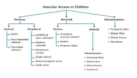
3.1.1 - Peripheral Intravenous Cannulation
Peripheral intravenous cannulation is common procedures for hospitalized patients which is still technically demanding procedure in neonates because their veins are small, fragile, and prone to collapse. As a result, multiple attempts are often required to achieve successful access. The first-attempt success rate for neonatal peripheral cannulation is relatively low, averaging around 45%. Some neonates may require several attempts, and reports describe up to 10–12 attempts in difficult cases, highlighting the procedural difficulty.
Techniques to Improve Success i) Strict aseptic precautions must be maintained during cannulation to reduce the risk of infection.
ii) Vein Visualization
- Transillumination: Frequently used in neonates to improve visualization of superficial veins.
- Ultrasound guidance: Increasingly employed in difficult cannulation cases to increase success rates.
iii) Stabilization of the Cannula- After insertion, the catheter should be secured with adhesive tape and a splint to minimize movement and reduce the risk of dislodgement. The insertion site should remain visible for monitoring.
iv)Pain Management- Non-pharmacological measures such as oral sucrose can help reduce procedural pain and distress.
v. Site Selection-The dorsum of the hand is the most commonly used site (approximately 40–42% of cases). Other potential sites include - Scalp veins, Foot veins, Forearm veins vi)Procedural Tips
- Limit attempts to three per operator before seeking assistance from a more experienced practitioner.
- Insert the cannula at an angle of approximately 10–30°.
- Ensure adequate limb immobilization during and after the procedure.[6], [7], [8], [9], [10], [11]
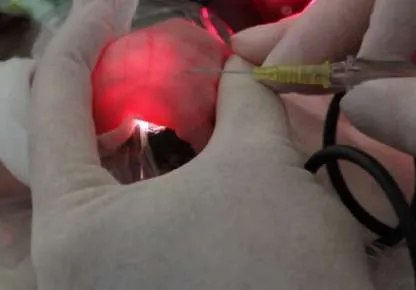
insertion using Venoscope
3.3.2 Umbilical Catheters
During neonatal period, one of the most frequently used central venous access devices is the umbilical venous catheter (UVC). In patients like preterm or critically ill infants who require intravenous administration of fluids, medications, and parenteral nutrition it can be easily placed and it is extremely useful. The catheter is inserted from umbilical vein which later in its course passes through the ductus venosus (DV) the tip should be located at the junction between the inferior vena cava (IVC) and the right atrium (RA).[12], [13], [14], [15], [16]
There are different types of catheters available based on material used they are Polyurethane, polyethylene and polyvinyl catheters. Amongst these Polyurethane catheters are preferred as they are less prone to bacterial colonization. These days Polyurethane UVCs that release antimicrobially active silver ions are available in the market. These catheters are potentially low risk of CLABSI, since they decrease both endoluminal and extraluminal colonization.[17]
3.3.2a Insertion Technique of Umbilical Catheter
Dunn formula is the most commonly used method for estimation of catheter length. Other formula used are Shukla and Ferrara, Verheij et al, Vali et al, Gupta et al. In emergency settings, an umbilical venous catheter (UVC) should be inserted only a short distance—about 2–4 cm just enough to allow blood to be freely aspirated. Advancing the catheter further at this stage can be dangerous, as medications are being infused directly into the liver, which may lead to hepatic injury. Normally, the catheter travels from the umbilical vein through the portal system and ductus venosus to reach the junction of the inferior vena cava and right atrium, but it can sometimes take an incorrect path into the liver. Simple techniques such as gently mobilizing the liver, positioning the baby on the right side, or applying mild upper abdominal pressure—especially under ultrasound guidance—can help guide the catheter along the correct route. Since umbilical line placement is a key skill expected of pediatric trainees, simulation-based training has become increasingly important, allowing learners to build confidence and competence without putting newborns at risk.
One of the most important advances in UVC placement is the growing use of real-time ultrasound or point-of-care ultrasound (POCUS). Unlike chest X-rays, which rely on indirect landmarks and may miss catheter misplacement, ultrasound allows clinicians to see both the vessel and the catheter tip directly during the procedure. Studies show that a significant number of catheters thought to be correctly positioned on X-ray need adjustment when ultrasound is used. Although ultrasound requires specific training, even short, focused teaching sessions have been shown to help doctors, residents, and neonatal nurse practitioners accurately identify catheter position quickly and confidently. As a result, real-time ultrasound-guided UVC insertion is emerging as a safer, more accurate, and cost-effective approach, with strong potential to become standard practice in neonatal intensive care units.[18]
| Reference | Study Population | Method Used | Formula / Result |
|---|---|---|---|
| Dunn[19] | 50 UV (necropsy) BW: 680–4027 g EG: 26–44 weeks | Anthropometric measurement | Shoulder–umbilicus distance |
| Shukla & Ferrara[20] | 39 UV, 4 UAC BW: 2037 ± 1077 g | Weight-based formula | Length (cm) = [(3 × BW in kg + 9) / 2 + 1] |
| Verheij et al.[21] | 143 UV (Shukla formula) 125 UV (revised formula) | Modified weight-based formula | Length (cm) = [(3 × BW in kg + 9) / 2] |
| Vail et al.[22] | 82 UV BW: 1311 ± 888 g EG: 23–27+6 weeks | External landmark measurement | Umbilicus–mid-xiphoid distance |
| Gupta et al.[23] | 170 UV BW: 490 ± 480 g EG: 24–41 weeks | External landmark formula | Length (cm) = Umbilical–nipple length − 1 |
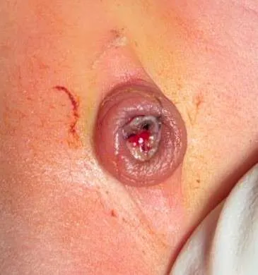
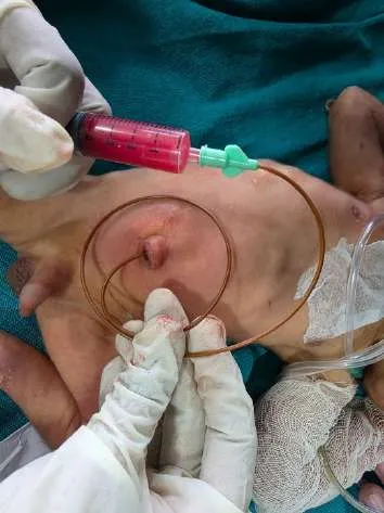
artery catheterization
3.3.3 Central Venous Access
Central venous catheters (CVCs), including peripherally inserted central catheters (PICCs), play a crucial role in the management of critically ill neonates in neonatal intensive care units (NICUs). These catheters provide reliable vascular access for the administration of parenteral nutrition, medications, hyperosmolar solutions, and for frequent blood sampling in premature or severely ill infants. Because peripheral venous access in neonates is often difficult and short-lived, central venous access becomes essential for prolonged therapy.
3.3.3a - Types of Central Venous Catheters
In neonates, central venous access is most commonly achieved using peripherally inserted central catheters (PICCs), also referred to as percutaneously inserted central venous catheters (PCVCs). These catheters are inserted through peripheral veins and advanced until the tip lies within a central vein. In situations where percutaneous access is unsuccessful or long-term access is required, tunneled catheters or surgically placed central venous lines may be used. Umbilical venous catheters (UVCs) are also commonly used in the early neonatal period Central venous catheters are indicated in neonates for:
- Administration of total parenteral nutrition (TPN)
- Delivery of hyperosmolar solutions (>900 mOsm/L)
- Infusion of medications with extreme pH (<5 or >9) such as vancomycin or phenytoin
- Long-term intravenous therapy
- Frequent blood sampling in critically ill neonates.[24], [25], [26]
3.3.3b - PICC (Peripherally inserted central venous catheter) Lines
Use of peripherally inserted central catheters (PICCs) started in the first decade of 1980. PICCs plays a vital role in improving the care quality among pediatric patients for long-term intravenous chemotherapeutics and antibiotics. They also have an essential role in neonates admitted to neonatal intensive care units (NICU) for a long time and receive parenteral nutrition and drugs. PICCs are a salient tool for the management of critically ill neonate and children, they have various advantages like ease of placement, fewer complications, shorter procedure time, a higher rate of patient satisfaction, and a more reliable form of intravenous (IV) access.[27]
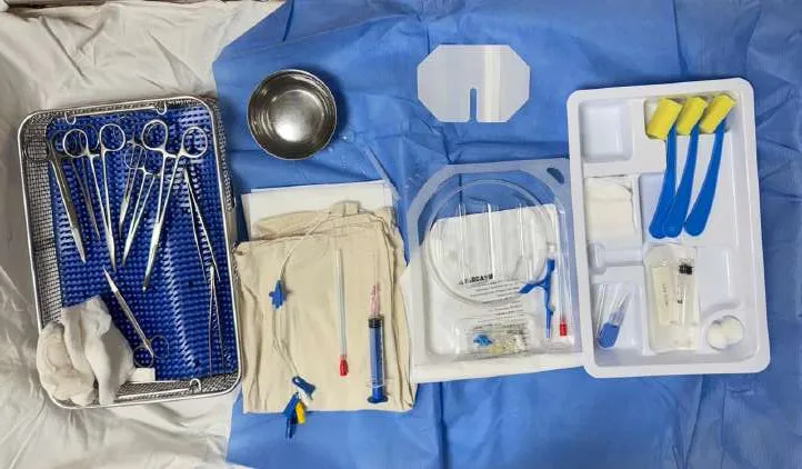
Peripherally inserted central catheters (PICCs) are available in 3 configurations - single-, double- , and triple-lumen and their sizes ranges from 28 G catheters for use in premature neonates to 7 Fr triple-lumen catheters. Traditionally, single-lumen catheters are described by gauge, while multi-lumen catheters by French (Fr) size. The size of the PICC to be used is determined by the size of the access vein and the therapy required and not simply by the age of the patient. In general, smaller catheters with the least number of lumens are associated with the fewest complications; however, very small catheters are more likely to become blocked. If blood sampling via the PICC is required, then at least a size 3 Fr will be needed. Any I.V. infusate can be given by a PICC, but their dwell time (the length of time the catheter remains in the vein) is typically <2 months and rarely >6 months. PICCs labelled as ‘power injectable’ can withstand high pressures, allowing I.V. contrast administration during CT scanning.[4]
Common intravenous External External Diameter French gauge
cannula needle gauge Diameter (mm) (mm)
24 0.7 - -
22 0.9 - -
20 1.1 1 3
18 1.3 1.34 4
17 1.5 - -
16 1.7 1.67 5
14 2.1 2 6
- - 2.3 7
Table 3 – External and internal diameter of PICC line
3.3.3c - Sites for PICCs insertion
Arms, legs, and scalp are commonly used insertion sites. Other possible sites for neonatal PICCs are the veins of the hands and feet. The basilic vein is larger and less tortuous than the cephalic vein. As the right basilic vein has a more direct route to central circulation it is preferred over the left basilic vein. The axillary vein is large and provides a direct route to the subclavian vein, however, while choosing axillary vein one should be careful not to cannulate axillary artery. Scalp veins are easily visualized but may become tortuous at the level of the ear and on entrance to the subclavian veins. In such cases moving the patient’s head to a midline position during insertion may facilitate catheter advancement to the central circulation. Leg veins offer multiple sites for neonatal PICC insertion, but advancing the catheter at the level of the femoral fold can be difficult. Femoral veins and jugular veins are best visualized and cannulated with ultrasound guidance.[28]
3.3.3d - PICC line insertion technique
Under all aseptic precautions, preparation of a fine-tipped introducer cannula (FTIC) is done to start the insertion. The PICC set consist of peelable introducer cannula which is partially peeled and needle is discarded. A 24-G peripheral venous cannula is passed through it fine-tipped introducer this peeling helps to make the fine-tipped introducer suitable for neonatal veins. An appropriate vein is the one with no prior puncture. The limb is sterilized, draped, immobilized, and a tourniquet applied to distend the vein. A small puncture is made which facilitate introducer entry, after this FTIC is advanced into the vein, until free blood return is seen. after intravascular placement is confirmed, the needle and 24-G cannula is removed and FTIC is left securely in the vein. Tourniquet is removed and a pre-flushed 2-Fr PICC is advanced through the FTIC to a predetermined length reaching the central vein. The introducer is then peeled away completely and removed, hemostasis is achieved with mild compression, and a small gauze tamponade and transparent sterile dressing are applied. The catheter tip position is confirmed by X-ray.[29]
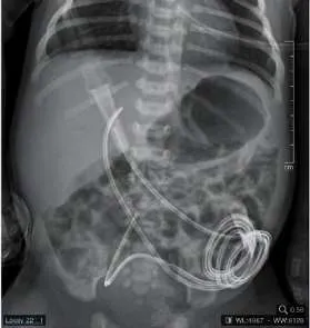
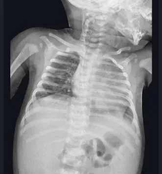
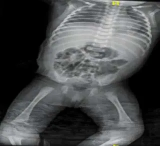
neonates
3.3.4 Tunneled Catheter
Tunneled CVCs end away from the venous access site and have a subcutaneous path. The duration of tunneled CVCs might range from weeks to months. Tunneled CVCs come in two main varieties: those that leave the skin as access catheters and those that terminate in a subcutaneous port. The subcutaneous port ends in a single or double port, depending on whether it has one or two lumens. This port is positioned beneath the skin and subcutaneous tissues on the chest wall. Tunnel catheter has variable tip and different lumen designs.[30], [31] The different tip design helps in maintaining patency and minimizing recirculation during dialysis.[32] Usual veins for tunnel catheter insertion re Right internal jugular vein, left internal jugular vein, Left subclavian vein and right subclavian vein.[33]
3.3.5 Intraosseous Access (IO)
IO has 91% success rate for establishing vascular access in critically ill newborns and is a safe, rapid, and effective procedure specially when conventional techniques fail.[34], [35], [36] A special hollow-bore needle is inserted through the bone cortex into the medullary space. It aids with the infusion of medications and fluids because neonates have relatively high vascular and cellular marrow as well as helps with blood sampling and investigations.[37], [38], [39] . Proximal tibia (the site may be located by placing a finger 1 cm below the tibial tuberosity and then sliding the finger 1 cm medially), the distal tibia (2 cm above the medial malleolus), and the distal femur are the preferable puncture sites in children for intraosseous access. Most commonly indicated puncture site is the proximal tibia because of presence of thin layer covering of skin on the anterior region of this bone and this site would not interfere with cardiopulmonary arrest procedures, such as chest compressions and invasive respiratory access for ventilation.[40] Contraindications for IO is Fracture of the target bone, previous infection at the site, and significant soft tissue injury.
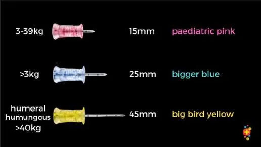
4. Complications
Most important complication associated with central line is (CLABSI Central-line associated bloodstream infection)CLABSI is defined by the U.S. Center for Disease Control and Prevention (CDC)/National Healthcare Safety Network (NHSN) for healthcare -associated infection surveillance as a primary bloodstream infection, e.g., recovery of a pathogen from a blood culture (on two separate occasions) in a patient of any age with a central line within 48 h before developing an infection that must not be related to an alternative cause. In infants ≤ 1 year of age, there is the additional requirement of having at least one of the following signs: fever (> 38 °C), hypothermia (< 36 °C), apnea, or bradycardia.[41], [42], [43], [44], [45]
Other Complications associated with umbilical vein catheterization and PICC line catheter are limb edema, line occlusion, malposition, leakage, sepsis, displacement, line thrombus, pleural effusion, pericardial effusion, line related complications, local inflammation, atrial thrombus, liver hemorrhage, limb ischemia.[46], [47]
Intraosseous access was found to have complications like misplacement in soft tissue, extravasation, healing deficiency and local swelling and rare complications like limb ischemia, tibial fracture.[48], [49]
In a systemic review done by Rebecca et. Al., it was found that peripherally inserted central venous catheter has 50% higher risk of dysfunction in children <1 years of age. When compared with PICC lines CVAD were associated with higher rates of thrombosis, approx. 60%, insertion failure, and infection. Though PIVs have lower risk of infection compared to CVAD, they have relatively higher risk of occlusion [50].
5.Surgical Cut-Down Techniques
When comparatively less invasive percutaneous methods like the Seldinger technique (percutaneous access), ultrasound-guided venous access, and intraosseous vascular access have failed, the venous cutdown technique is a surgical procedure to provide venous access. Venous cutdown is a life-saving option because percutaneous access can be challenging for some patient populations (pediatric patients with small, nonpalpable veins, patients in hypovolemic shock with collapsed veins, patients with peripheral vascular disease with altered vascular anatomy).[51], [52]
6. Ultrasound In Vascular Access
Ultrasound plays a major role in neonatal vascular access. It is useful in both arterial as well as venous access. According to various small sectorial probe, 7–8 MHz, is used for deep veins as it gives preferably better visualization. Micro-convex probes, 7–8.5 MHz. linear “hockey stick” probe 10–14 MHz, gives better visualization of tissue up to a depth of 3–5 cm. The image plane is oriented along the vessel's 'short axis' or along the ‘long axis with the use of B mode. Both vein and artery appear similar with anechoic lumen. Generally, arteries due to its thick wall are less compressible which helps to distinguish arteries from veins. Real-time ultrasound guidance using sterile technique is the most effective method, allowing direct visualization of needle entry into the vein, confirmation of blood aspiration, and safe guidewire placement by a single operator
7. Summary
Vascular access is a critical component of neonatal care, particularly in neonatal intensive care units (NICUs), where reliable access is required for administration of fluids, medications, parenteral nutrition, and blood sampling. Due to the small size and fragility of neonatal vessels, establishing and maintaining vascular access can be technically challenging and requires careful selection of the appropriate technique.
Peripheral venous access is the most commonly used method and is usually obtained from veins on the dorsum of the hand, foot, scalp, or antecubital region. It is simple, minimally invasive, and suitable for short-term therapy. However, peripheral lines often have a limited dwell time and may require frequent replacement.
For intermediate or long-term therapy, central venous access is frequently required. This may be achieved through peripherally inserted central catheters (PICCs) or centrally inserted catheters placed in the internal jugular, subclavian, or femoral veins. These lines allow administration of hyperosmolar solutions such as total parenteral nutrition and enable repeated blood sampling. Umbilical venous catheters are commonly used during the early neonatal period and provide rapid and reliable central access.
In emergency situations where intravenous access cannot be quickly established, intraosseous access provides a rapid alternative for the administration of fluids and medications. Arterial access, typically through the radial or posterior tibial artery, is used for continuous blood pressure monitoring and arterial blood gas analysis.
Each vascular access method has specific advantages, limitations, and potential complications, including infection, thrombosis, catheter occlusion, and mechanical injury. Therefore, careful technique, strict aseptic precautions, and regular monitoring are essential to minimize complications and ensure safe use.
Overall, the choice of vascular access in neonates depends on the clinical condition of the infant, duration of therapy, type of infuscate, and availability of expertise, with the goal of achieving reliable access while minimizing risks.
BIBILOGRAPHY – [1] V. Scott-Warren MBChB FRCA and R. B. Morley FRCA, “Paediatric vascular access,” British Journal of Anaesthesia Education , vol. 15, pp. 199 –206, 2015, doi: 10.1093/bjaceaccp/mku050.
[2] “Vascular Access | Pediatric Surgery NaT.” Accessed: Nov. 05, 2025. [Online]. Available:
https://www.pedsurglibrary.com/apsa/view/Pediatric-Surgery- NaT/829031/all/Vascular_Access [3] “Clinical Practice Guidelines : Intravenous access - Peripheral.” Accessed: Nov. 10, 2025.
[Online]. Available:
https://www.rch.org.au/clinicalguide/guideline_index/Intravenous_access_Peripheral/ [4] V. L. Scott-Warren and R. B. Morley, “Paediatric vascular access,” BJA Educ., vol. 15, no.
4, pp. 199–206, Aug. 2015, doi: 10.1093/bjaceaccp/mku050.
[5] V. M. Naik, S. Shyam Prasad Mantha, and B. K. Rayani, “Vascular access in children,” Indian J. Anaesth., vol. 63, no. 9, p. 737, Sep. 2019, doi: 10.4103/IJA.IJA_489_19. [6] Y. Okada et al., “Effectiveness of ultrasound-guided peripheral arterial cannulation in neonates, including very low birth weight infants who are conventionally difficult-to-cannulate: a case series,” European Journal of Pediatrics 2023 183:1, vol. 183, no. 1, pp. 509–512, Nov. 2023, doi: 10.1007/s00431-023-05320-6.
[7] C. Casal-Guisande et al., “Peripheral Vascular Access in Infants: Is Ultrasound-Guided Cannulation More Effective than the Conventional Approach? A Systematic Review,” Medicina 2025, Vol. 61, Page 1321, vol. 61, no. 8, p. 1321, Jul. 2025, doi: 10.3390/medicina61081321.
[8] S. Hall et al., “Recurrent peripheral intravenous catheterization in neonates: A case series,” Nurs. Crit. Care, vol. 30, no. 5, Sep. 2025, doi: 10.1111/nicc.13232.
[9] S. R. Tandale, N. Dave, M. Garasia, S. Patil, and S. Parelkar, “A Study of Morbidity and Cost of Peripheral Venous Cannulation in Neonates Admitted to Paediatric Surgical Intensive Care Unit,” J. Clin. Diagn. Res., vol. 11, no. 3, p. UC08, Mar. 2017, doi: 10.7860/JCDR/2017/23600.9570.
[10] “Intravenous Cannulation in neonates.” Accessed: Mar. 07, 2026. [Online]. Available:
https://www.starship.org.nz/guidelines/intravenous-cannulation-in-neonates/ [11] P. C. Parmar, B. Shah, and A. Mehta, “Improving peripheral vein cannulation in neonates with skin transillumination: a prospective observational study by a neonatal fellow at a tertiary care centre,” Int. J. Contemp. Pediatrics, vol. 12, no. 4, pp. 626–631, Mar. 2025, doi: 10.18203/2349-3291.ijcp20250769.
[12] J. Anderson, D. Leonard, D. A. V. Braner, S. Lai, and K. Tegtmeyer, “Videos in clinical medicine. Umbilical vascular catheterization,” N. Engl. J. Med., vol. 359, no. 15, p. e18, Oct. 2008, doi: 10.1056/NEJMVCM0800666.
[13] M. C. Hermansen and G. M. Hermansen, “Intravascular catheter complications in the neonatal intensive care unit,” Clin. Perinatol., vol. 32, no. 1, pp. 141–156, 2005, doi: 10.1016/j.clp.2004.11.005.
[14] R. E. Campbell, “Roentgenologic features of umbilical vascular catheterization in the newborn,” Am. J. Roentgenol. Radium Ther. Nucl. Med., vol. 112, no. 1, pp. 68–76, 1971, doi: 10.2214/AJR.112.1.68.
[15] A. E. Oestreich, “Umbilical vein catheterization--appropriate and inappropriate placement,” Pediatr. Radiol., vol. 40, no. 12, pp. 1941–1949, Dec. 2010, doi: 10.1007/S00247-010- 1840-2.
[16] “Gorski: Umbilical cathethers - Google Scholar.” Accessed: Dec. 03, 2025. [Online].
Available:
https://scholar.google.com/scholar_lookup?author=L+Gorski&author=L+Hadaway&autho r=ME+Hagle&author=M+McGoldrick&author=M+Orr&author=D+Doellman+&publicati on_year=2021&title=Umbilical+cathethers&journal=Infusion+Therapy+Standards+of+Pr actice&pages=S90-3 [17] J. S. Garland et al., “Cohort study of the pathogenesis and molecular epidemiology of catheter-related bloodstream infection in neonates with peripherally inserted central venous catheters,” Infect. Control Hosp. Epidemiol., vol. 29, no. 3, pp. 243–249, Mar. 2008, doi: 10.1086/526439.
[18] M. Wagner et al., “Umbilical Venous Catheter Update: A Narrative Review Including Ultrasound and Training,” 2022, doi: 10.3389/fped.2021.774705.
[19] P. M. Dunn, “Localization of the umbilical catheter by post-mortem measurement.,” Arch.
Dis. Child., vol. 41, no. 215, pp. 69–75, Feb. 1966, doi: 10.1136/ADC.41.215.69. [20] H. Shukla and A. Ferrara, “Rapid estimation of insertional length of umbilical catheters in newborns,” Am. J. Dis. Child., vol. 140, no. 8, pp. 786 –788, 1986, doi: 10.1001/ARCHPEDI.1986.02140220068034.
[21] G. H. Verheij, A. B. Te Pas, V. E. H. J. Smits-Wintjens, A. Šràmek, F. J. Walther, and E.
Lopriore, “Revised formula to determine the insertion length of umbilical vein catheters,” Eur. J. Pediatr., vol. 172, no. 8, pp. 1011–1015, Aug. 2013, doi: 10.1007/S00431-013-1981- Z.
[22] P. Vali, S. E. Fleming, and J. H. Kim, “Determination of umbilical catheter placement using anatomic landmarks,” Neonatology, vol. 98, no. 4, pp. 381–386, Nov. 2010, doi: 10.1159/000316918.
[23] A. O. Gupta, M. R. Peesay, and J. Ramasethu, “Simple measurements to place umbilical catheters using surface anatomy,” J. Perinatol., vol. 35, no. 7, pp. 476–480, Jul. 2015, doi: 10.1038/JP.2014.239.
[24] D. W. Cartwright, “Central venous lines in neonates: a study of 2186 catheters,” Arch. Dis.
Child. Fetal Neonatal Ed., vol. 89, no. 6, pp. F504 –F508, Nov. 2004, doi: 10.1136/adc.2004.049189.
[25] J. Retzmann et al., “Central venous catheters in very low birthweight infants: results from a national survey,” Journal of Hospital Infection, vol. 165, pp. 73–80, Nov. 2025, doi: 10.1016/j.jhin.2025.07.027.
[26] U. Trieschmann, U. ten F. Cate, and N. Sreeram, “Central venous catheters in children and neonates – what is important?,” Images Paediatr. Cardiol., vol. 9, no. 4, p. 1, Oct. 2007, Accessed: Mar. 07, 2026. [Online]. Available:
https://pmc.ncbi.nlm.nih.gov/articles/PMC3232582/ [27] G. Bahoush, P. Salajegheh, A. M. Anari, A. Eshghi, B. H. Aski, and A. Asghar, “A review of peripherally inserted central catheters and various types of vascular access in very small children and pediatric patients and their potential complications,” 2021, doi: 10.25122/jml- 2020-0011.
[28] “(PDF) PICC Placement in the Neonate.” Accessed: Nov. 15, 2025. [Online]. Available:
https://www.researchgate.net/publication/343163018_PICC_Placement_in_the_Neonate [29] I. Uygun, “Peripherally inserted central catheter in neonates: A safe and easy insertion technique,” J. Pediatr. Surg., vol. 51, no. 1, pp. 188 –191, Jan. 2016, doi: 10.1016/j.jpedsurg.2015.08.008.
[30] M. Gallieni, I. Brenna, F. Brunini, N. Mezzina, S. Pasho, and A. Giordano, “Dialysis central venous catheter types and performance,” J. Vasc. Access, vol. 15 Suppl 7, no. SUPPL. 7, 2014, doi: 10.5301/JVA.5000262. [31] “Advances in Tunneled Central Venous Catheters for Dialysis: Design and Performance - Ash - 2008 - Seminars in Dialysis - Wiley Online Library.” Accessed: Dec. 02, 2025. [Online]. Available: https://onlinelibrary.wiley.com/doi/10.1111/j.1525 - 139X.2008.00494.x [32] K. Boubes, A. Shaikh, Z. Alsauskas, and A. Dwyer, “New Directions in Ensuring Catheter Safety,” Adv. Chronic Kidney Dis., vol. 27, no. 3, pp. 228–235, May 2020, doi: 10.1053/J.ACKD.2020.02.004.
[33] R. Nourzaie et al., “Atypical use of PICC as centrally inserted central catheter in infants and neonates: Report of a 10-year experience,” Journal of Vascular Access, vol. 24, no. 3, pp. 409–415, May 2023, doi: 10.1177/11297298211034308.
[34] G. Strandberg, A. Larsson, M. Lipcsey, and M. Eriksson, “Comparison of Intraosseous, Arterial, and Venous Blood Sampling for Laboratory Analysis in Hemorrhagic Shock,” Clin. Lab., vol. 65, no. 7, pp. 1169–1177, 2019, doi: 10.7754/CLIN.LAB.2019.181214. [35] M. Jousi, J. Björkman, and J. Nurmi, “Point-of-care analyses of blood samples from intraosseous access in pre-hospital critical care,” Acta Anaesthesiol. Scand., vol. 63, no. 10, pp. 1419–1425, Nov. 2019, doi: 10.1111/AAS.13443.
[36] H. Ellemunter, B. Simma, R. Trawöger, and H. Maurer, “Intraosseous lines in preterm and full term neonates,” Arch. Dis. Child. Fetal Neonatal Ed., vol. 80, no. 1, 1999, doi: 10.1136/FN.80.1.F74.
[37] “Vascular access through the intraosseous route in pediatric emergencies”.
[38] M. L. Ling et al., “APSIC guide for prevention of Central Line Associated Bloodstream Infections (CLABSI),” Antimicrob. Resist. Infect. Control, vol. 5, no. 1, p. 16, May 2016, doi: 10.1186/S13756-016-0116-5.
[39] T. Bell and N. P. O’Grady, “Prevention of Central Line–Associated Bloodstream Infections,” Infect. Dis. Clin. North Am., vol. 31, no. 3, pp. 551–559, Sep. 2017, doi: 10.1016/j.idc.2017.05.007.
[40] “BSI | PSC | NHSN | CDC.” Accessed: Dec. 07, 2025. [Online]. Available:
https://www.cdc.gov/nhsn/psc/bsi/index.html [41] M. O. Wright, S. G. Decker, K. Allen-Bridson, J. N. Hebden, and D. Leaptrot, “Healthcare-associated infections studies project: An American Journal of Infection Control and National Healthcare Safety Network data quality collaboration: Location mapping,” Am. J. Infect. Control, vol. 46, no. 5, pp. 577–578, May 2018, doi: 10.1016/j.ajic.2017.12.012. [42] H. J. Cho and H. K. Cho, “Central line-associated bloodstream infections in neonates,” Korean J. Pediatr., vol. 62, no. 3, pp. 79–84, Dec. 2018, doi: 10.3345/KJP.2018.07003. [43] S. Arun et al., “Comparison of UVC with PICC Line for Reducing Central Line Associated Blood Stream Infections in Preterm Neonates with Birth Weight < 1250g: An Open-Label Randomized Controlled Trial”.
[44] S. Avanzini et al., “Brachiocephalic vein for percutaneous ultrasound-guided central line positioning in children: A 20-month preliminary experience with 109 procedures,” Pediatr. Blood Cancer, vol. 64, no. 2, pp. 330–335, Feb. 2017, doi: 10.1002/PBC.26202. [45] E. Schwindt et al., “Intraosseous access in neonates is feasible and safe – An analysis of a prospective nationwide surveillance study in Germany,” Front. Pediatr., vol. 10, p. 952632, Jul. 2022, doi: 10.3389/FPED.2022.952632/BIBTEX.
[46] A. Scrivens et al., “Use of Intraosseous Needles in Neonates: A Systematic Review,” Neonatology, vol. 116, no. 4, pp. 305–314, Nov. 2019, doi: 10.1159/000502212. [47] R. S. Paterson et al., “Selection and insertion of vascular access devices in pediatrics: A systematic review,” Pediatrics, vol. 145, pp. S243 –S268, Jun. 2020, doi: 10.1542/PEDS.2019-3474H.
[48] R. Taghizadeh and P. M. Gilbert, “Long saphenous venous cutdown revisited,” Burns, vol.
32, no. 2, pp. 267–268, Mar. 2006, doi: 10.1016/j.burns.2005.10.022.
[49] S. Chappell, G. M. Vilke, T. C. Chan, R. A. Harrigan, and J. W. Ufberg, “Peripheral venous cutdown,” Journal of Emergency Medicine, vol. 31, no. 4, pp. 411–416, Nov. 2006, doi: 10.1016/j.jemermed.2006.05.026.
[50] I. Cole, C. Glass, H. J. Norton, and V. Tayal, “Ultrasound measurements of the saphenous vein in the pediatric emergency department population with comparison to i.v. Catheter size,” Journal of Emergency Medicine, vol. 43, no. 1, pp. 87–92, Jul. 2012, doi: 10.1016/j.jemermed.2011.06.034.
[51] M. Pittiruti, M. G. Annetta, and V. D’andrea, “Point-of-care ultrasound for vascular access in neonates and children,” European Journal of Pediatrics 2023 183:3, vol. 183, no. 3, pp. 1073–1078, Dec. 2023, doi: 10.1007/S00431-023-05378-2.
[52] I. Ikonomidis et al., “Academic Editors: Peripheral Vascular Access in Infants: Is Ultrasound-Guided Cannulation More Effective than the Conventional Approach? A Systematic Review,” 2025, doi: 10.3390/medicina61081321.